Why Histology Biomedical Scientists are so Important within the NHS
Cancer diagnosis is a complex process which relies on a wide range of healthcare workers. Included in this process are biomedical scientists who work within the histology laboratories. Though not as publicly recognised as patient-facing workers, histologists play a pivotal role in identifying cancerous tissues, which then helps to shape the course of treatment for patients.
The Process of Histology in Cancer Diagnosis
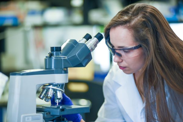
Working as a recruiter in Biomedical Science, I have learnt about the process of Histology itself, along with how it contributes to cancer diagnosis. The primary task of a Biomedical Scientist in Histology is to prepare and examine tissue samples under a microscope, which will then enable doctors to make informed diagnoses. These tissue samples can come from biopsies, surgeries, or other medical procedures, and they provide invaluable insight into the cellular makeup of organs, tissues, and tumours. Their ability to view and understand these minute structures at a cellular level is crucial in diagnosing diseases, monitoring treatment progress, and guiding therapeutic decisions.
So, there are a few steps that Biomedical Scientists need to take when checking a sample for cancerous tissue, the process of cancer diagnosis through histology generally follows several key steps:
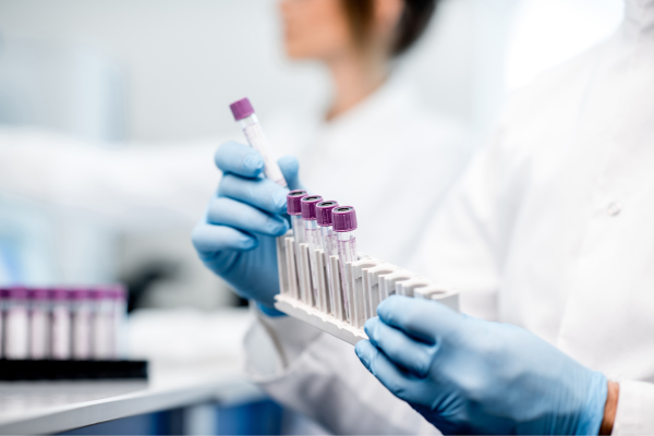
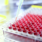
- Tissue Collection (Biopsy): The tissue sample is collected from the patient during a biopsy or surgery. This could involve removing part or all lesions, masses, or tumours. Once the tissue is obtained, it is immediately sent to the histology lab.
- Fixation and Processing: The collected tissue is first "fixed," which preserves the cellular structures by preventing decay. After fixation, the tissue is processed through a series of steps that dehydrate and embed the sample in paraffin wax. This makes the tissue firm enough to be sliced into thin sections.
- Tissue Sectioning: The thin sections of the tissue are cut using an instrument called a microtome. These slices are so thin that they can be mounted on glass slides, creating a sample that can be stained and examined under a microscope. Some experienced histologists can confidentially cut up to 50 blocks per hour!
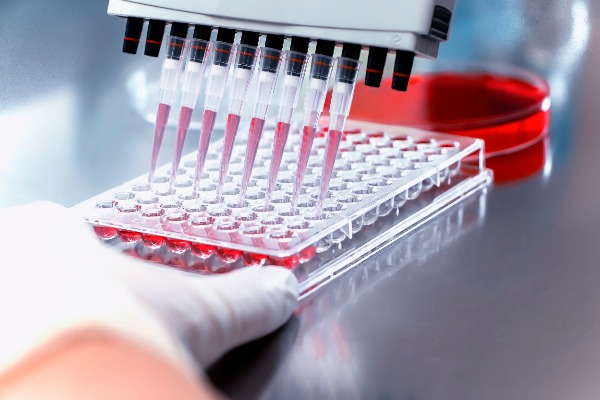
- Staining: Once the tissue is mounted on a slide, a special stain is applied to make the different cellular structures visible and create a contrast that allows biomedical scientists to differentiate between normal and abnormal tissue patterns.
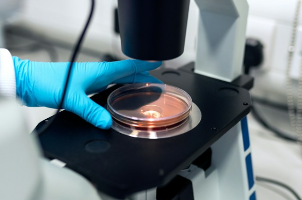
- Microscopic Examination: The slide is then ready for microscopic examination which is when experts check the slides to accurately identify cellular abnormalities. The slide is then examined to look for signs of cancer, such as changes in the shape, size, and/or arrangement of cells.
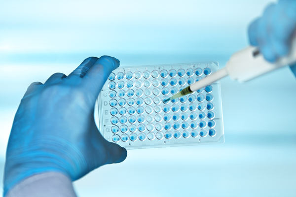
- Immunohistochemistry (IHC) and Advanced Techniques: In some cases, additional staining techniques, such as immunohistochemistry (IHC), are required to provide more detailed information. IHC involves using antibodies to detect specific proteins in the cells, helping to classify the cancer more precisely (e.g., identifying the type of cancer and its aggressiveness).
How Biomedical Scientists Contribute to Cancer Diagnosis
Biomedical Scientists are responsible for ensuring the quality and accuracy of the tissue samples, which will lead to making the correct diagnosis. Poorly prepared slides can lead to misinterpretation, delays in diagnosis, or incorrect treatments so the quality of the slides is important to ensure the correct abnormalities are highlighted, which may indicate cancer.
Moreover, biomedical scientists may encounter variations in tissue types, sizes, and conditions, requiring them to adjust their techniques accordingly. They must be detail-oriented and knowledgeable about the specific requirements for different types of tissues and tumours. Their expertise allows for consistent, high-quality preparation of specimens.
Biomedical Scientists can help diagnose a variety of cancers, including:
- Breast cancer: They help differentiate between benign and malignant tumours, and aid in identifying hormone receptor status, which influences treatment decisions.
- Lung cancer: They help distinguish between different types of lung cancer, such as small cell or non-small cell lung cancer.
- Colon cancer: They examine polyps and lesions to determine if they are cancerous or precancerous.
- Prostate cancer: They play a key role in grading prostate cancer using the Gleason score, which informs prognosis and treatment options.
The Unsung Heroes Behind Accurate Cancer Diagnosis
The work that Biomedical Scientists do behind the scenes is crucial for determining the stage of the disease, planning the course of treatment, and providing prognostic information. Through their expertise in tissue preparation, staining, and microscopic examination, they provide the essential foundation for accurate diagnoses. Their meticulous work is critical in identifying cancer at its earliest stages, guiding treatment, and ultimately saving lives. While they may not be the face of cancer treatment, histologists are indispensable to the process of diagnosing and managing this complex disease.
As highlighted by Maisie Harris, Senior Recruitment Consultant and author of this article, the expertise of Histology Biomedical Scientists is indispensable to the process of diagnosing and managing this complex disease.
Get in Touch with Maisie Harris
If you’d like to discuss the insights shared in this article or explore placements in Histology, feel free to reach out to Maisie Harris. She’s here to answer your questions and help you navigate your career in Biomedical Science.
Latest Biomedical Science Jobs
We currently have a variety of locum and permanent vacancies across the UK and Ireland